$67.5
$129.6
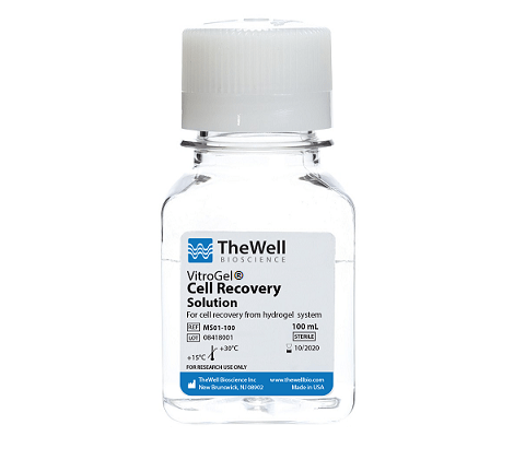
VitroGel Cell Recovery Solution – Cell Harvesting VitroGel® Cell Recovery Solution is an non-enzymatic cell harvesting solution to recover cells/organoids cultured in 2D, 3D efficiently and safely in 20 minutes. VitroGel Cell Recovery Solution is room temperature stable, has a neutral pH, and works at 37°C operating temperature. The solution can maintain high cell viability during the recovery process. Harvested cells can be sub-culture in both 2D and 3D cultures. The VitroGel Cell Recovery Solution can be used before or after the fixation and stained preparation of hydrogel specimens to ensure high-quality downstream data analysis. Easy cell recovery from hydrogel in 20 minutes. Learn how cell harvesting works Specifications Size100 mL FormulationEnzyme-free UseHarvest cells from VitroGel hydrogel while maintaining high cell viability.Use before or after sample fixation and stained preparation for imaging or downstream data analysis Processing Time15-20 min DownstreamRecovered cells can be sub-culture in both 2D and 3D culture pHNeutral StorageAmbient Temperature (15-30°C) Stability60 months from date of manufacture Protocols / Resources Product Documentation VitroGel Cell Recovery Solution Sale Sheet Product Data Sheet Frequently Asked Questions Material Safety Data Sheet (MSDS) Protocol VitroGel Cell Recovery Solution – Protocol Video Protocols & Demonstrations VIDEO PROTOCOL TIP Cell Recovery from 3D & 2D VitroGel Hydrogel TECHNICAL TIPS Technical Tips KEEP SOLUTION WARM: It is important to keep the cell recovery solution and the mixture warm at 37°C during the whole process. The warm temperature is essential to accelerate molecular exchanges to release the ionic molecules from the solid hydrogel, which can transform into a soft hydrogel. APPLY MECHANICAL FORCE: The mechanical force, such as rocking or shaking the centrifuge tube or using a serological pipette to mix the hydrogel with the cell recovery solution helps to transform the hydrogel into the liquid state. DILUTION: Adding the cell recovery solution at a volume of 10X or higher than the hydrogel maintains the dissolved hydrogel in a liquid state. CENTRIFUGE AT ROOM TEMPERATURE Application Notes APPLICATION NOTE Quick and Efficient Organoid/Cell Recovery from Animal-Based Extracellular Matrix APPLICATION NOTE Long-Term Neuron Culture Maturation in 3D Hydrogel Constructs APPLICATION NOTE 3D Cell Culture of Human Colon Cancer Cells (HCT116) on VitroGel® System APPLICATION NOTE 3D Cell Culture of Human Pancreatic Cancer Cells (PANC-1) on VitroGel® System Data and References Figure 1. Fast hydrogel dissolved in VitroGel Cell Recovery Solution. A. Hydrogel before adding to recovery solution; B-F. Time 0 to 15 min after adding hydrogel to recovery solution (at 37 °C, 20 rpm). /wp-content/uploads/2019/08/VitroGel-Cell-Recovery-Solution-Time-Lapse.mp4 Figure 2 The VitroGel Cell Recovery Solution maintains high cell viability A. Cell viability of OP9, U87-MG and PANC-1 cells after adding to recovery solution at time 0, 15, 30, 60 and 120 min. Cells maintain over 95% cell viability after suspending in VitroGel cell recovery solution for 2 hr.; B. PANC-1 cells growth on 2D well plate before transfer to cell recovery solution; C. PANC-1 cells suspended in cell recovery solution for 24 hours then re-culture on 2D well plate for 5 days. Cells has been successful re-culture after suspend in cell recovery solution for 24 hours. Figure 3. Cell viability of 3D cultured PANC-1 cells after recovering from hydrogel. (Method 1: add whole gel into cell recovery solution. Method 2: using pipette to break gel into small piece before adding into cell recovery solution. Average: the average cell viability of method 1 and method 2.) Figure 4. Cells can be sub-culture in both 2D and 3D culture after recovery. A. PANC-1 cells growth on 3D hydrogel before harvested by VitroGel cell recovery solution; B. PANC-1 cells have been harvested from 3D hydrogel by using VitroGel cell recovery solution and subculture on the surface of hydrogel (day 2); C. PANC-1 cells have been harvested from 3D hydrogel by using the cell recovery solution and 3D subculture in the hydrogel system again (day 2). References/Publications McKinnon, B., Duempelmann, L., Skrabalova, J., Subramaniam, S., Taylor, L., Atluri, S., Chauquet, S., Montgomery, G., Tanaka, K., Ramm, S., Luu, J., Simpson, K., Shah, S., Amoako, A., Mueller, M., & Nirgianakis, K. (2026). Dienogest interrupts the chemokine signalling axis in endometriotic lesions with resistant signatures, providing novel opportunities for personalised treatment. https://doi.org/10.21203/rs.3.rs-8096027/v1 Li, F., Xu, T., Sun, R., He, Y., Lin, J.-F., Chen, H., Wang, J., Chen, J., Chen, P., Guo, Q., Yang, Q., Cai, W., Li, C., Zeng, M., Cao, J., Fan, J., Huang, X., Wang, Q., & Zhang, Q. (2025). CircPPP1CB subtype, hsa_circ_0007439, promotes nasopharyngeal carcinoma progression by upregulating KRT1. Discover Oncology, 16(1), 2031–2031. https://doi.org/10.1007/s12672-025-03888-z Fuego, D. M., Devkota, I., Bonomo, Z. L., Li, Y., Zhang, X., Dondeti, M. F., Donnarumma, F., Matsakas, A., Patterson, A. L., Williams, C. C., Vourekas, A., Fu, X., Bondioli, K. R., & Simintiras, C. A. (2025). Oviduct fluid metabolic regulation of embryonic genome methylation. https://doi.org/10.1101/2025.06.13.659599 Lee, H. J., Lau, L. N., Sidhu, S. K., Park, J.-Y., & Yeo, I.-S. L. (2025). Three-Dimensional Hydrogel Culture Reveals Novel Differentiation Potential of Human Bone Marrow-Derived Stem Cells. Prosthesis, 7(3), 52. https://doi.org/10.3390/prosthesis7030052 Li, F., Song, L., He, Y., Chen, P., Wang, J., Zeng, M., Li, C., Chen, J., Chen, H., Guo, Q., Fan, J., Huang, X., Wang, Q., & Zhang, Q. (2025). FLT1-enriched extracellular vesicles induce a positive feedback loop between nasopharyngeal carcinoma cells and endothelial cells to promote angiogenesis and tumour metastasis. Oncogene. https://doi.org/10.1038/s41388-025-03389-x Liu, X., Zhou, Z., Lu, X., Zhong, H., He, R., Feng, Z., & Guan, R. (2024). Protective mechanism of quercetin nanoliposomes on hydrogen peroxide-induced oxidative damage in 3D Caco-2 cell model. Journal of Functional Foods, 124, 106650. https://doi.org/10.1016/j.jff.2024.106650 Gabusi, E., Lenzi, E., Manferdini, C., Dolzani, P., Columbaro, M., Saleh, Y., & Lisignoli, G. (2022). Autophagy Is a Crucial Path in Chondrogenesis of Adipose-Derived Mesenchymal Stromal Cells Laden in Hydrogel. Gels , 8(12),766. https://www.mdpi.com/2310-2861/8/12/766 De Donato, M., Babini, G., Mozzetti, S., Buttarelli, M., Ciucci, A., Arduini, G., De Rosa, M. C., Scambia, G., & Gallo, D. (2020). KLF7: a new candidate biomarker and therapeutic target for high-grade serous ovarian cancer. Journal of Experimental & Clinical Cancer Research, 39(1). https://doi.org/10.1186/s13046-020-01775-9 Lan, T., Guo, J., Bai, X., Huang, Z., Wei, Z., Du, G., Yan, G., Weng, L., & Yi, X. (2020). RGD-modified injectable hydrogel maintains islet beta-cell survival and function. Journal of Applied Biomaterials & Functional Materials, 18, 228080002096347. https://doi.org/10.1177/2280800020963473 Powell K. Adding depth to cell culture. Science, 356(6333), 96–98. https://doi.org/10.1126/science.356.6333.96 References to all VitroGel hydrogels >
Ready-To-Use Hydrogels